Main Content
Visual Enhancement
Info and registration 655 6244 Open Mon 8.00–16.00 Tues.–Thurs. 8.00–18.00 Fri 8.00–16.00
Info and registration 655 6244 Open Mon 8.00–16.00 Tues.–Thurs. 8.00–18.00 Fri 8.00–16.00
Glaucoma is a condition of the eye that can lead to the destruction of nerve fibers and an irreversible deterioration of vision. Glaucoma can have a variety of causes, but in many cases, it is caused by an elevated intraocular pressure.
Due to damage to the nerve fibers, the vision of a glaucoma patient deteriorates. As the disease progresses, the narrowing of the visual field becomes more noticeable.

Scientific studies show that hereditary predisposition plays a major role in glaucoma. Therefore, it is important that the offspring of glaucoma patients (who are over 40 years of age) have their eyes thoroughly examined once a year.
If glaucoma is detected and treated, it is usually possible to stop the disease from getting worse.
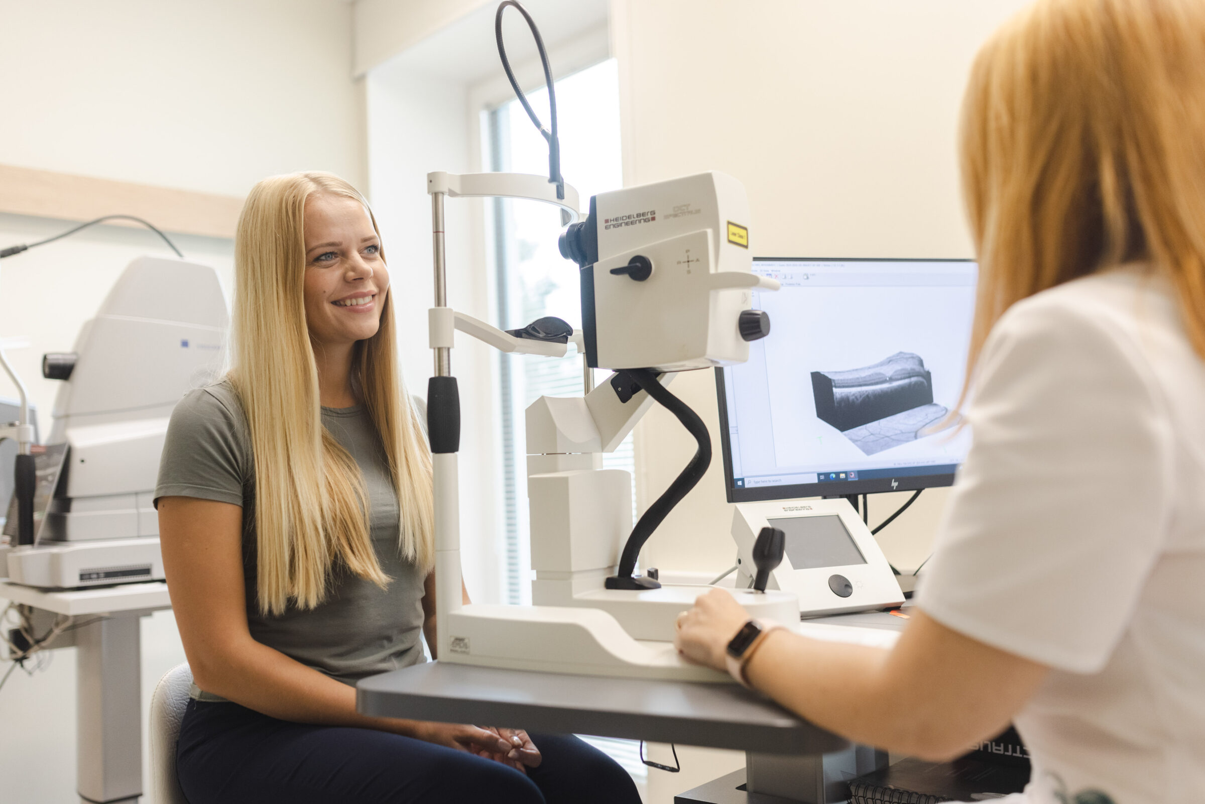
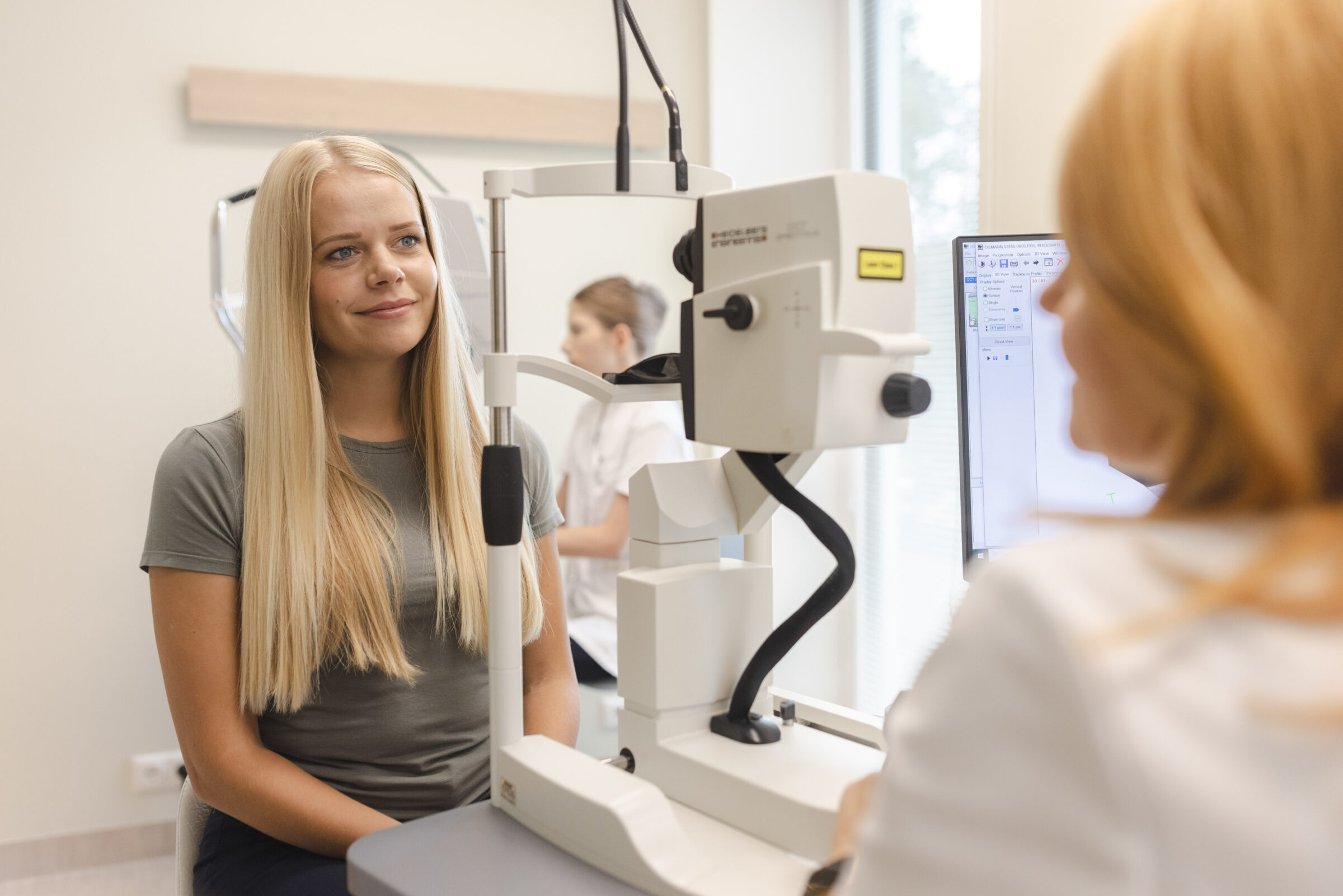
Optical coherence tomography (OCT) is a novel imaging technique. It automatically analyses the various structures in the eye in which glaucoma may have caused changes. These are the optic nerve disc, the layer of nerve fibers, and the layer of ganglion cells. Because these structures are tiny, it is usually difficult to detect disease-related changes with conventional methods. The Spectralis® OCT scan makes this possible for your ophthalmologist.
A visual perimetry test can be used to assess the sensitivity of the eye and the extent of peripheral vision. Damage to the nerve fibers in the ocular fundus caused by glaucoma is mainly studied, but it can also be implemented for other vision problems. There are several different examinations of the visual field. The consultant will determine the most suitable one for you.
The Procedure
is simple. The patient places their chin on a chin rest and will observe a series of flashing lights on the oval screen. The flashes may have different levels of brightness and appear at various points in the visual field. Upon noticing each flash, the patient must press the button, and the device will record what is seen. The device checks each point repeatedly, so when the patient makes an error, the device can correct it.
The test usually takes about 5–7 minutes, but this may vary.
How to prepare for the test?
Contact lenses may be worn during the test, but if subsequent tests or a consultation are scheduled, it is advised to wear glasses. If you use reading glasses, please bring them with you.
The Spectralis® OCT test allows disease-related changes to be detected quite early – before the patient begins to perceive them due to the narrowing of the field of vision. This way, treatment can be initiated early and effectively.
If you have already been diagnosed with glaucoma, the Spectralis® OCT scan allows us to monitor the course of your disease precisely and to plan your treatment optimally.