Main Content
Visual Enhancement
Info and registration 655 6244 Open Mon 8.00–16.00 Tues.–Thurs. 8.00–18.00 Fri 8.00–16.00
Info and registration 655 6244 Open Mon 8.00–16.00 Tues.–Thurs. 8.00–18.00 Fri 8.00–16.00
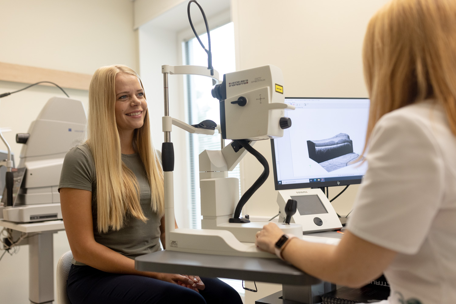
Spectralis® is an image processing platform that can be used for diagnostic and treatment decision making. The device provides excellent contrast and high-resolution images of both the front and back of the eye.
HEIDELBERG ENGINEERING SPECTRALIS enables us to examine the eye in record speed and many levels, discovering primary changes already at the micro level and imaging different cross-sections of the layers of the eye:
Spectralis® gives us the opportunity to observe various layers of the eye without having to dilate the pupils of the patient, which is vital among the following patient groups:
Spectralis® is equipped with ground breaking technology called Blue Laser. This the most effective technology for photographing the fundus, as all the energy is focused on one specific wavelength (so far, a wide spectrum of light has been used for photographing the fundus). As a result, the light flash disturbs the patient much less and does not have an unpleasant effect.
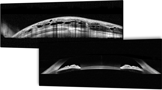
provides an overview of the corneal layers and anterior chamber. The width of the cornea and the measure of the anterior chamber angle provide relevant additional information in the diagnosis of glaucoma.
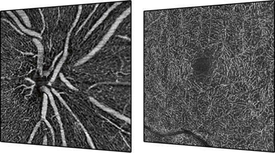
is the latest module allowing us to examine blood vessels without injecting a contrast agent. It is particularly suitable for allergic patients to avoid possible complications of fluorescein angiography.
The new generation of eye fundus computed tomography has the advantage of faster imaging speed and sharper image quality, which highlights retinal layers and their changes. Pupil size will not be affected during this examination.
is the new generation of glaucoma diagnostics. Measuring the width of the optic nerve disc, retinal nerve fibers, and the layer of ganglion cells is important in diagnosing glaucoma and monitoring the course of the disease.
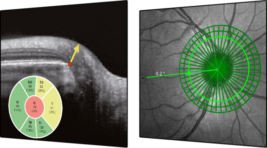
The instrument is able to measure the exact same location as in the previous study in subsequent examinations and to perform a comparative analysis of the anatomical structures on the basis of the information obtained. In addition, patient data can be compared to a statistical average.
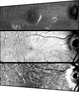
uses three laser wavelengths in parallel to detect different structures at different depths in the retina. The method allows us to visualize changes that cannot be seen with normal fundus examination or images. The described module can be used in imaging when the patient has cataracts or nystagmus.
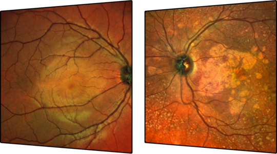
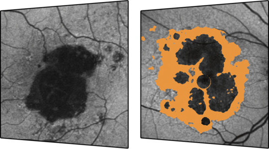
indicates blue laser autofluorescence images. The non-invasive laser method for imaging of the eye fundus, reveals traces of retinal metabolic stress using lipofuscin as an indicator. The method highlights changes in retinal pigment-epithelial cells, such as age-related macular degeneration of the eye fundus and its hereditary changes.
Multicolor, OCT, OCT Angiography, and BluePeak Spectralis modules provide a wider imaging range than older devices. Thus, they allow more peripheral areas of the fundus to be photographed and documented.