Info and registration 655 6244 Open Mon 8.00–16.00 Tues.–Thurs. 8.00–18.00 Fri 8.00–16.00
Info and registration 655 6244 Open Mon 8.00–16.00 Tues.–Thurs. 8.00–18.00 Fri 8.00–16.00
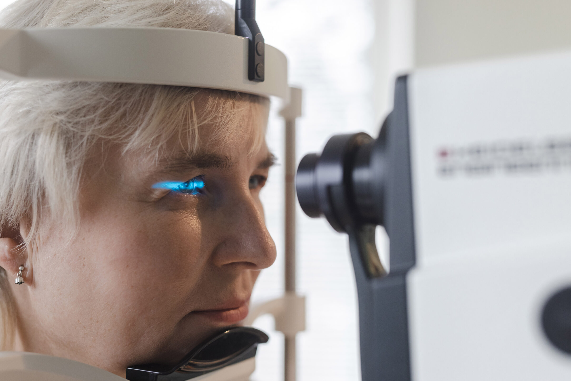
Fluorescence angiography or fluorescein angiography is a diagnostic procedure in which the fundus is imaged and the retina and blood vessels are mapped. The procedure can detect a variety of pathological processes, such as vascular disease, bruising, edema, and neoplasms.
OCT-A is an additional module for Optical Coherent Tomography (OCT) that displays retinal and choroidal microvessels.
OCT-A technology uses a laser beam that reflects back from red blood cells moving in the blood vessel. If there is no movement or blood flow in the blood vessels, an image of the blood vessels cannot be obtained. The method is used in particular for the diagnosis of diabetic retinopathy, thrombosis, and the wet form of AMD.
The examination is not painful or uncomfortable, and it only takes a little longer than regular OCT. The contrast agent is not injected, which makes this form of angiography significantly safer than conventional fluorescein angiography (FAG/FA).
Unfortunately, the OCT-A method is not always sufficient to diagnose diseases, and a FAG must still be performed.
Fluorescence angiography, or fluorescein angiography of the ocular fundus, is a diagnostic procedure in which the fundus is imaged and the retina and blood vessels are mapped. The procedure can detect a variety of pathological processes, such as vascular disease-indicative changes, bruising, edema, and neoplasms.

The dye, fluorescein, is injected into a vein in the patient’s arm, which enters the blood vessels of the eye fundus through the bloodstream and highlights possible lesions in the fundus.
Fluorescein is a bright yellow dye that turns the skin slightly yellow for a few hours. The dye is excreted by the kidneys, which turns the color of the urine bright yellow for 24 to 36 hours.
The following side effects have occasionally been reported regarding the usage of fluorescein: fatigue, nausea, vomiting, headache, stomach irritation, absent-mindedness, fainting, itching, and rash. There have been reports of life-threatening allergic reactions (anaphylactic shock) or difficulty breathing due to an allergic reaction in the bronchi.

Leakage of the dye from the blood vessels to the basal tissues of the skin has been noted if the venous cannula is dislodged or ruptured in the vein wall during injection. However, these complications are uncommon. Should this occur, we recommend using a compression wrap at the injection site for a few days.
Intravenous fluorescein is not usually administered to pregnant or breast-feeding mothers, although there is no scientific reasoning indicating that it could harm the unborn baby or affect the breast milk.
Please allow up to one and a half hours (including preparation) for the ocular fundus angiography.